Mycobacterium ulcerans - Buruli ulcer
Overview | Project page
Within the past decade, the incidence of Buruli ulcer has dramatically increased in several countries in sub-Saharan Africa, Australia, Asia, Mexico, and Peru, leading the WHO to declare this disease a global health threat in 2004. Despite being the third most common mycobacterial disease in immunocompetent humans after tuberculosis and leprosy, Buruli ulcer is considered a “neglected tropical disease” by the WHO and does not garner the attention given to other infectious diseases. The WHO estimates that the incidence of Buruli ulcer will surpass that of leprosy and could become more problematic than tuberculosis in some African regions.
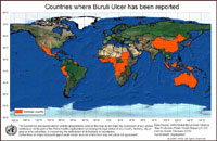 (click map for larger view)
Mycobacterium ulcerans
Mycobacterium ulcerans, the microorganism that causes Buruli ulcer, is a slow-growing environmental mycobacterium of which the natural reservoir is unknown. Human transmission is believed to occur via skin transmission by direct inoculation or an insect vector. Most individuals infected with M. ulcerans initially develop a small, painless, pre-ulcerative skin nodule with larger areas of indurated skin and edema. As the disease progresses over one to two months, the infected skin begins to ulcerate with characteristic necrosis of the subcutaneous fatty tissues, deeply undermined edges, and vascular blockage. These necrotic ulcers can lead to very extensive skin loss, damage to nerves, blood vessels, and appendages, and deformity and disability, particularly in children.
Currently, no vaccine is available for the prevention of Buruli ulcer. Although antibiotic treatment has been shown to be effective in vitro and in animal models, success in the clinical environment has produced variable results, especially in the case of advanced ulcerative disease. Accordingly, surgical excision, combined with antibiotic therapy, prevails as an accepted remedy for these difficult-to-treat infections.
Warning: The blue boxes are concealing graphic images that may be disturbing
to some viewers. If you would like to reveal the images, simply put your mouse
on the blue box.

back to top
Treatment of Buruli ulcer with clay minerals
Documented use of two clay minerals as a therapeutic treatment for the necrotizing Mycobacterium ulcerans skin infection, Buruli ulcer, suggests that specific natural mineral products have significant beneficial effects on wound healing. In 2001, a French humanitarian working in the Ivory Coast of Africa began treating children with Buruli ulcer with natural clay minerals. Within days of initiating treatment with clay poultices, the therapeutic properties of the clay minerals were demonstrated, with the initiation of rapid, non-surgical debridement of the destroyed tissue. Extended treatment with the clay minerals resulted in continued debridement of the ulcer, tissue regeneration, and wound healing. After several months of daily clay applications, the Buruli ulcer wounds healed with soft, supple scarring and return of normal motor function.
back to top